Prevention is better than cure
A superb study and a highly relevant one...
The technology, infrastructure and the medical services have decreased the fatality in stroke. while this does lead to independence for ADL and professional activities also in some cases, many stay dependent, putting a burden on the family and the society.
What we have to do is not just to improve the post-event medical facilities, but to improve the pre-event medical system so that stroke does not happen. That would be our true victory....and sadly no business for me, an interventional neuroradiologist.
Remarkable Decline in Ischemic Stroke Mortality Is not Matched by Changes in Incidence [Original Contributions]:
Background and Purpose—
In Western Europe, mortality from ischemic stroke (IS) has declined over several decades. Age–sex-specific IS mortality, IS incidence, 30-day case fatality, and 1-year mortality after hospital admission are essential for explaining recent trends in IS mortality in the new millennium.
Methods—
Data for all IS deaths (1980–2010) in the Netherlands were grouped by year, sex, and age. A joinpoint regression was fitted to detect points in time at which significant changes in the trends occur. By linking nationwide registers, a cohort of patients first admitted for IS between 1997 and 2005 was constructed and age–sex-specific 30-day case fatality and 1-year mortality were computed. IS incidence (admitted IS patients and out-of-hospital IS deaths) was computed by age and sex. Mann–Kendall tests were used for trend evaluation.
Results—
IS mortality declined continuously between1980 and 2000 with an attenuation of decline in the 1990s in some of the age–sex groups. A remarkable decline in IS mortality after 2000 was observed in all age–sex groups, except for young men. An improved decline in 30-day case fatality and in 1-year mortality was also observed in almost all age–sex groups. In contrast, IS incidence remained stable between 1997 and 2005 or even increased slightly.
Conclusions—
The recent remarkable decline in IS mortality was not matched by a decline in the number of incident nonfatal IS events. This is worrying, because IS is already a leading cause of adult disability, claiming a heavy human and economic burden. Prevention of IS is therefore now of the greatest importance.
Is Intra-Arterial Thrombolysis Beneficial for M2 Occlusions? Subgroup Analysis of the PROACT-II Trial [Brief Report]
A very interesting article.....I have thought of doing IAT for such 'distal MCA' occlusions often but somehow always found noninvasive management better -either IVT or medical management as indicated...
The problem for me is the smaller infarct volume and relatively preserved clinical status of such patients, at least in my practice, and the complication rate associated with the IAT.
But of course 50+ % success rate is reasonably good....but in our center we have fairly good clinical outcomes in such patient with IVT...
I am still not impressed in spite of this article...
Is Intra-Arterial Thrombolysis Beneficial for M2 Occlusions? Subgroup Analysis of the PROACT-II Trial [Brief Report]:
Rahme, R et al, Stroke ASAP Dec 2012
Background and Purpose—
The role of endovascular therapy for acute M2 trunk occlusions is debatable. Through a subgroup analysis of Prolyse in Acute Cerebral Thromboembolism-II, we compared outcomes of M2 occlusions in treatment and control arms.
Methods—
Solitary M2 occlusions were identified from the Prolyse in Acute Cerebral Thromboembolism-II database. Primary endpoints were successful angiographic reperfusion (TICI 2–3) at 120 minutes and functional independence (mRS 0–2) at 90 days.
Results—
Forty-four patients with solitary M2 occlusions, 30 in the treatment arm and 14 in the control arm, were identified. Successful reperfusion (TICI 2–3) was achieved in 53.6% and 16.7% of patients in the treatment and control arms, respectively (P=0.04). A favorable clinical outcome (mRS 0–2) was observed in 53.3% and 28.6%, respectively (P=0.19). Baseline characteristics were similar between the 2 groups.
Conclusions—
Intra-arterial thrombolysis may lead to a 3-fold increase in the rate of early reperfusion of solitary M2 occlusions and could potentially double the chance of a favorable functional outcome at 90 days.
Clinical Trial Registration—
This trial was not registered because enrollment began before July 1, 2005.
Strategic coil placement at the mid-body of Pcom artery aneurysm
Aneurysm come in all shape and sizes and at all locations.
This one was in a 42 years old female patient with Grade I SAH and a ruptured right PcomA aneurysm.
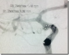
The anatomy was odd with a bulbous, rather blister like proximal part, then a narrowing, then the body, again a narrowing then a teat like portion. The Pcom, of course, had to arise from the aneurysm; specifically it came at the site of first narrowing mentioned.
The aneurysm was directed laterally and posteriorly and had a curved structure rather.
The proximal bulbous portion measured 2.67 mm in diameter with equal neck and the distal narrower portions 2 mm.
So, I used an Echelon and Xpedion –ten system- to access the aneurysm.
Then i was in a fix as to how to go about fixing the aneurysm.
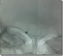
I put in a 2x6 3D AXIUM in the mid part of the aneurysm beyond the Pcom origin. The coil loops did try to go in the distal teat but somehow the entire coil could be fit in there.
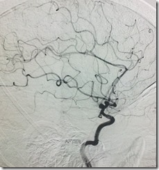
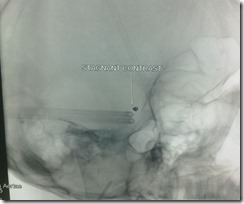
Angio showed complete cessation of flow within that portion, with contrast stasis in the teat and the Pcom stayed patent.
However in the native images, still some space appeared within the coil mesh, so I took an AXIUM 2x4 Helix coil and pushed in.
The loops went into the first coil and then started to come into the proximal portion. Somehow, the loops in this part stayed horizontal thus restructuring the inflow zone and contrast flowing into the pcom.
But the last loop could not be fit in and kept pushing the microcatheter tip into the arterial lumen.
So I left the procedure at that point and put my hands up.
The patient is fine, and we all are happy, but do not know what this anueurysm was ot how best to treat it.
My surgeon too told he would have clipped in the midpart so the result would have been same, or rather bad as this looked to me like an infundibulum which had ruptured and the distal portion to me was nothing but a pseudoaneurysm.
I am keeping my fingers crossed. let’s see….
Preoperative partial embolization of thoracic hemangioblastoma
One place for all your notes and information
Heparin dosing is associated with diffusion weighted imaging lesion load following aneurysm coiling
Heparin dosing is associated with diffusion weighted imaging lesion load following aneurysm coiling: Background and purpose
Diffusion weighted imaging (DWI) may be used to evaluate post-coiling ischemia. Heparinization protocols for cerebral aneurysm coiling procedures differ among operators and centers, with little literature surrounding its effect on DWI lesions. The goal of this study was to determine which factors, including heparinization protocols, may affect DWI lesion load post-coiling.
Materials and methods
A review of 135 coiling procedures over 5 years at our centre was performed. Procedural data including length of procedure, number of coils used, stent or balloon assistance and operators were collected. Procedures were either assigned as using a bolus dose (>2000 U at any one time) or small aliquots of heparin (≤2000 U). Postprocedure DWI was reviewed and lesions were classified as small (< 5mm), medium (5–10 mm) or large (>10 mm). The cases were then classified into group 1 (≤5 small lesions) or group 2 (>5 small lesions or ≥1 medium or large lesion). Multivariate regression of the procedural variables for the two groups was calculated. A p value of <0.05 was considered significant.
Results
There were 78 procedures in group 1 and 57 procedures in group 2. Patients who received small aliquots (n=37) versus boluses of heparin (n=98) intraprocedurally had significantly greater frequency and size of DWI lesions (p=0.03). None of the other procedural variables was found to impact on lesion load.
Conclusions
More substantial DWI lesions were associated with small aliquots of heparin dosage compared with bolus doses. Heparin boluses should be preferentially administered during aneurysm coiling.
The hyperdense vessel sign on CT predicts successful recanalization with the...
Sent to you by subbu via Google Reader:
Background
The success of mechanical clot retrieval for acute ischemic stroke may be influenced by the characteristics of the occlusive thrombus. The thrombus can be partly characterized by CT, as the hyperdense vessel sign (HVS) suggests erythrocyte-rich clot whereas fibrin-rich clot may be isodense. We hypothesized that the physical clot characteristics that determine CT density may also determine likelihood of retrieval with the Merci device.
MethodsWe reviewed all acute stroke cases initially imaged with non-contrast CT before attempted Merci clot retrieval at a single center between 2004 and 2010. Each CT was blindly assessed for the presence or absence of the HVS, and post-retrieval angiograms were blindly assessed for reperfusion using the TICI scale.
ResultsOf 67 patients analyzed (mean age 69; median NIHSS 19; 61% female), the HVS was seen in 42, and no HVS was present in 25. Successful recanalization was achieved in 79% of patients with the HVS (33/42), but in only 36% (9/25) of patients without HVS (p=0.001). The HVS was the only significant predictor of recanalization while accounting for age, treatment with IV-tPA, clot location, stroke etiology, time to treatment, and number of retrieval attempts.
ConclusionThe HVS in acute ischemic stroke was strongly predictive of successful recanalization using the Merci device. The HVS may indicate thrombi that are less adhesive compared with isodense clots that are more resistant to mechanical retrieval. The absence of HVS on pre-treatment CT may thus suggest the need for a more aggressive or alternative therapeutic approach.
Things you can do from here:
- Subscribe to Journal of NeuroInterventional Surgery Online First using Google Reader
- Get started using Google Reader to easily keep up with all your favorite sites
Clinical Significance of Impaired Cerebrovascular Autoregulation After Sever...
Sent to you by subbu via Google Reader:
Background and Purpose—
The purpose of this study was to investigate the relationship between cerebrovascular autoregulation and outcome after aneurysmal subarachnoid hemorrhage.
Methods—In a prospective observational study, 80 patients after severe subarachnoid hemorrhage were continuously monitored for cerebral perfusion pressure and partial pressure of brain tissue oxygen for an average of 7.9 days (range, 1.9–14.9 days). Autoregulation was assessed using the index of brain tissue oxygen pressure reactivity (ORx), a moving correlation coefficient between cerebral perfusion pressure and partial pressure of brain tissue oxygen. High ORx indicates impaired autoregulation; low ORx signifies intact autoregulation. Outcome was determined at 6 months and dichotomized into favorable (Glasgow Outcome Scale 4–5) and unfavorable outcome (Glasgow Outcome Scale 1–3).
Results—Twenty-four patients had a favorable and 56 an unfavorable outcome. In a univariate analysis, there were significant differences in autoregulation (ORx 0.19±0.10 versus 0.37±0.11, P<0.001, for favorable versus unfavorable outcome, respectively), age (44.1±11.0 years versus 54.2±12.1 years, P=0.001), occurrence of delayed cerebral infarction (8% versus 46%, P<0.001), use of coiling (25% versus 54%, P=0.02), partial pressure of brain tissue oxygen (24.9±6.6 mmüHg versus 21.8±6.3 mmüHg, P=0.048), and Fisher grade (P=0.03). In a multivariate analysis, ORx (P<0.001) and age (P=0.003) retained an independent predictive value for outcome. ORx correlated with Glasgow Outcome Scale (r=–0.70, P<0.001).
Conclusions—The status of cerebrovascular autoregulation might be an important pathophysiological factor in the disease process after subarachnoid hemorrhage, because impaired autoregulation was independently associated with an unfavorable outcome.
Things you can do from here:
- Subscribe to Stroke ASAP using Google Reader
- Get started using Google Reader to easily keep up with all your favorite sites
Endovascular Treatment of Intracranial Unruptured Aneurysms: A Systematic Re...
Sent to you by subbu via Google Reader:
Purpose:
To report subgroup analyses of an updated systematic review on endovascular treatment of intracranial unruptured aneurysms (UAs); to compare types of embolic agents, adjunct techniques, and newer devices; and to identify potential risk factors for poor outcomes.
Materials and Methods:Meta-Analysis of Observational Studies in Epidemiology and Preferred Reporting Items for Systematic Reviews and Meta-Analyses guidelines were used to prepare this article, and the literature was searched with PubMed and with EMBASE and Cochrane databases. Six eligibility criteria (procedural complications rates; at least 10 patients; saccular, nondissecting UAs; original study published in English or French between January 2003 and July 2011; methodological quality score > 6 [modified Strengthening and Reporting of Observational Studies in Epidemiology criteria]; a study published in a peer-reviewed journal) were used. End points included procedural mortality and unfavorable outcomes (death or modified Rankin Scale, Glasgow Outcome Scale, or World Federation of Neurosurgeons Scale at 1 month scores, all > 2). A fixed-effects model (Mantel-Haenszel) was used for pooled estimates of mortality and unfavorable outcomes; a random-effects model (DerSimonian-Laird) was used in case of heterogeneity.
Results:Ninety-seven studies with 7172 patients (26 studies published July 2008 through July 2011) were included. Sixty-nine (1.8%) of 7034 patients died (fixed-effect weighted average; 99% confidence interval [CI]: 1.4%, 2.4%; Q value, 55.0; I2 = 0%). Unfavorable outcomes, including death, occurred in 4.7% (242 of 6941) of patients (99% CI: 3.8, 5.7; Q value, 128.3; I2 = 26.8%). Patients treated after 2004 had better outcomes (unfavorable outcome, 3.1; 99% CI: 2.4, 4.0) than patients treated during 2001–2003 (unfavorable outcome, 4.7%; 99% CI: 3.6%, 6.1%; P = .01) or in 2000 and before (unfavorable outcome, 5.6%; 99% CI: 4.7%, 6.6%; P < .001). Significantly higher risk was associated with liquid embolic agents (8.1%; 99% CI: 4.7%, 13.7%) versus simple coil placement (4.9%; 99% CI: 3.8%, 6.3%; P = .002). Unfavorable outcomes occurred in 11.5% (99% CI: 4.9%, 24.6%) of patients treated with flow diversion.
Conclusion:Procedure-related poor outcomes occurred (4.7% of patients), risks decreased, and liquid embolic agents and flow diversion were associated with higher risks.
©RSNA, 2012
Supplemental material:http://radiology.rsna.org/lookup/suppl/doi:10.1148/radiol.12112114/-/DC1
Things you can do from here:
- Subscribe to Radiology current issue using Google Reader
- Get started using Google Reader to easily keep up with all your favorite sites
Closed-Cell Stent for Coil Embolization of Intracranial Aneurysms: Clinical ...
Sent to you by subbu via Google Reader:
BACKGROUND AND PURPOSE:
Recanalization is observed in 20–40% of endovascularly treated intracranial aneurysms. To further reduce the recanalization and expand endovascular treatment, we evaluated the safety and efficacy of closed-cell SACE.
MATERIALS AND METHODS:Between 2007 and 2010, 147 consecutive patients (110 women; mean age, 54 years) presenting at 2 centers with 161 wide-neck ruptured and unruptured aneurysms were treated by using SACE. Inclusion criteria were wide-neck aneurysms (>4 mm or a dome/neck ratio ≤2). Clinical outcomes were assessed by the mRS score at baseline, discharge, and follow-up. Aneurysm occlusion was assessed on angiograms by using the RS immediately after SACE and at follow-up.
RESULTS:Eighteen aneurysms (11%) were treated following rupture. Procedure-related mortality and permanent neurologic deficits occurred in 2 (1.4%) and 5 patients (3.4%), respectively. In total, 7 patients (4.8%) died, including 2 with reruptures. Of the 140 surviving patients, 113 (80.7%) patients with 120 aneurysms were available for follow-up neurologic examination at a mean of 11.8 months. An increase in mRS score from admission to follow-up by 1, 2, or 3 points was seen in 7 (6.9%), 1 (1%), and 2 (2%) patients, respectively. Follow-up angiography was performed in 120 aneurysms at a mean of 11.9 months. Recanalization occurred in 12 aneurysms (10%), requiring retreatment in 7 (5.8%). Moderate in-stent stenosis was seen in 1 (0.8%), which remained asymptomatic.
CONCLUSIONS:This series adds to the evidence demonstrating the safety and effectiveness of SACE in the treatment of intracranial aneurysms. However, SACE of ruptured aneurysms and premature termination of antiplatelet treatment are associated with increased morbidity and mortality.
Things you can do from here:
- Subscribe to Publication Preview using Google Reader
- Get started using Google Reader to easily keep up with all your favorite sites
Guidelines for the Management of Aneurysmal Subarachnoid Hemorrhage: A Guide...
Sent to you by subbu via Google Reader:
Purpose—
The aim of this guideline is to present current and comprehensive recommendations for the diagnosis and treatment of aneurysmal subarachnoid hemorrhage (aSAH).
Methods—A formal literature search of MEDLINE (November 1, 2006, through May 1, 2010) was performed. Data were synthesized with the use of evidence tables. Writing group members met by teleconference to discuss data-derived recommendations. The American Heart Association Stroke Council's Levels of Evidence grading algorithm was used to grade each recommendation. The guideline draft was reviewed by 7 expert peer reviewers and by the members of the Stroke Council Leadership and Manuscript Oversight Committees. It is intended that this guideline be fully updated every 3 years.
Results—Evidence-based guidelines are presented for the care of patients presenting with aSAH. The focus of the guideline was subdivided into incidence, risk factors, prevention, natural history and outcome, diagnosis, prevention of rebleeding, surgical and endovascular repair of ruptured aneurysms, systems of care, anesthetic management during repair, management of vasospasm and delayed cerebral ischemia, management of hydrocephalus, management of seizures, and management of medical complications.
Conclusions—aSAH is a serious medical condition in which outcome can be dramatically impacted by early, aggressive, expert care. The guidelines offer a framework for goal-directed treatment of the patient with aSAH.
Things you can do from here:
- Subscribe to Stroke current issue using Google Reader
- Get started using Google Reader to easily keep up with all your favorite sites
Single-Center Experience of Cerebral Artery Thrombectomy Using the TREVO Dev...
Sent to you by subbu via Google Reader:
Background and Purpose—
We sought to explore the safety and efficacy of the new TREVO stent-like retriever in consecutive patients with acute stroke.
Methods—We conducted a prospective, single-center study of 60 patients (mean age, 71.3 years; male 47%) with stroke lasting <8 hours in the anterior circulation (n=54) or <12 hours in the vertebrobasilar circulation (n=6) treated if CT perfusion/CT angiography confirmed a large artery occlusion, ruled out a malignant profile, or showed target mismatch if symptoms >4.5 hours. Successful recanalization (Thrombolysis In Cerebral Infarction 2b–3), good outcome (modified Rankin Scale score 0–2) and mortality at Day 90, device-related complications, and symptomatic hemorrhage (parenchymal hematoma Type 1 or parenchymal hematoma Type 2 and National Institutes of Health Stroke Scale score increment ≥4 points) were prospectively assessed.
Results—Median (interquartile range) National Institutes of Health Stroke Scale score on admission was 18 (12–22). The median (interquartile range) time from stroke onset to groin puncture was 210 (173–296) minutes. Successful revascularization was obtained in 44 (73.3%) of the cases when only the TREVO device was used and in 52 (86.7%) when other devices or additional intra-arterial tissue-type plasminogen activator were also required. The median time (interquartile range) of the procedure was 80 (45–114) minutes. Good outcome was achieved in 27 (45%) of the patients and the mortality rate was 28.3%. Seven patients (11.7%) presented a symptomatic intracranial hemorrhage. No other major complications were detected.
Conclusions—The TREVO device was reasonably safe and effective in patients with severe stroke. These results support further investigation of the TREVO device in multicentric registries and randomized clinical trials.
Things you can do from here:
- Subscribe to Stroke current issue using Google Reader
- Get started using Google Reader to easily keep up with all your favorite sites
Eligibility for Intravenous Recombinant Tissue-Type Plasminogen Activator Wi...
Sent to you by subbu via Google Reader:
Background and Purpose—
The publication of the European Cooperative Acute Stroke Study (ECASS III) expanded the treatment time to thrombolysis for acute ischemic stroke from 3 to 4.5 hours from symptom onset. The impact of the expanded time window on treatment rates has not been comprehensively evaluated in a population-based study.
Methods—All patients with an ischemic stroke presenting to an emergency department during calendar year 2005 in the 17 hospitals that compromise the large 1.3 million Greater Cincinnati/Northern Kentucky population were included in the analysis. Criteria for exclusion from thrombolytic therapy are analyzed retrospectively for both the standard and expanded timeframes with varying door-to-needle times.
Results—During the study period, 1838 ischemic strokes presenting to an emergency department were identified. A small proportion of them arrived in the expanded time window (3.4%) compared with the standard time window (22%). Only 0.5% of those who arrived in this timeframe met eligibility criteria for thrombolysis compared with 5.9% using standard eligibility criteria in the standard timeframe. These results did not vary significantly by repeated analysis varying the door-to-needle time or the expanded time window's exclusion criteria.
Conclusions—In reality, the expanded time window for thrombolysis in acute ischemic stroke benefits few patients. If we are to improve recombinant tissue-type plasminogen activator administration rates, our focus should be on improving stroke awareness, transport to facilities with ability to administer thrombolysis, and familiarity of physicians with acute stroke treatment guidelines.
Things you can do from here:
- Subscribe to Stroke current issue using Google Reader
- Get started using Google Reader to easily keep up with all your favorite sites
Venous thrombosis presenting as subarachnoid hemorrhage
- 49 yrs, M
- Seizures on the morning of admission
- Vitals stable
- No focal deficit
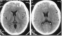
- A CT done at admission showed SAH with flocculent surface hematoma in the left anterior frontal region…significantly there was no blood in the basal cisterns
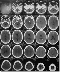
- NCCT at admission
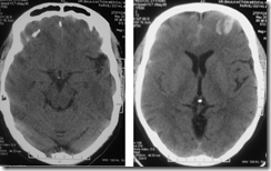

- Enlarged images of the NCCT
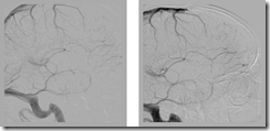
- A cerebral DSA was done which showed thrombosis of anterior part of the superior sagittal sinus, partial thrombosis of the inferior sagittal sinus with hypoplastic left transverse and sigmoid sinuses
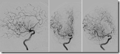

- Right ICA angiogram
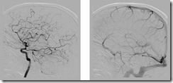
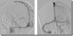
- Left ICA angiogram

- Left and right vertebral angiogram
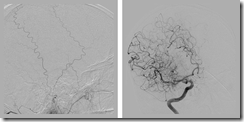
- Right middle meningeal had anomalous origin from right petrous ICA
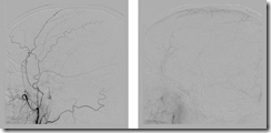
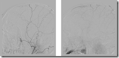
- Left ECA angiogram showed blush from the middle meningeal artery in the region of the thrombosed superior sagittal sinus
Patient was put on heparin after this
CLOTBUST sonothrombolysis of acute stroke
Unassisted coiling of a wide necked aneurysm
Here is an example, wherein a wide neck Acom aneurysm incorporating one of the A2 segments, was coiled well without use of any balloon/stent.
Long-Term Clinical and Imaging Follow-Up of Complex Intracranial Aneurysms Treated by Endovascular Parent Vessel Occlusion
Long-Term Clinical and Imaging Follow-Up of Complex Intracranial Aneurysms Treated by Endovascular Parent Vessel Occlusion
- From the Department of Neurosurgery, Neurovascular & Stroke Programs (C.C.M.), Yale University School of Medicine, New Haven, Connecticut; Division of Neuroradiology, Department of Medical Imaging (Z.K., K.G.t.B., R.A.W.), Toronto Western Hospital and the University of Toronto, Toronto, Ontario, Canada.
- Please address correspondence to Charles C. Matouk, Department of Neurosurgery, Neurovascular & Stroke Programs, Yale University School of Medicine, 333 Cedar St, TMP402, New Haven, CT, 06510; e-mail: charles.matouk@yale.edu
Abstract
Abbreviations
- BTO
- balloon test occlusion
- ECIC
- extracranial-intracranial
- PCA
- posterior cerebral artery
- PVO
- parent vessel occlusion
- VA
- vertebral artery
- VB
- vertebrobasilar











A.jpg)
A.jpg)
A.jpg)
A.jpg)


